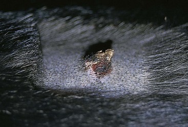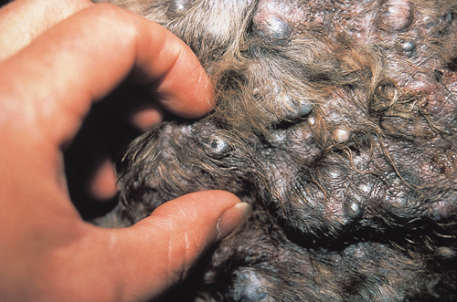
In this study the immunohistochemical expression of Hsp27 Hsp72 and Hsp73 was evaluated in normal canine skin 14 intracutaneous cornifying epitheliomas ICE 10 well-differentiated and 5 moderately differentiated squamous cell carcinomas SCC. He was castrated early at 6 months owing to an un-descended testicle.

All tumors were diagnosed by histologic examination.
Cornifying epitheliomas dog. The clinical histopathologic and behavioral features of 25 cases of intracutaneous cornifying epithelioma ICE in the dog were reviewed. A typical ICE consisted of a keratin-filled crypt in the dermis and subcutis that opened to the skin surface. Most of these tumors occurred on the back neck sides of the thorax and the shoulders.
Unknown but breed predispositions are recorded. Solitary or multiple keratin-filled cysts in dermis with demonstrable pore on surface. Well delineated and benign.
Clinical problems due to mass effect and with secondary inflammation after. Cornifying means looking like a horny substance and epitheliomas is a medical word for a skin tumor that can be either benign or cancerous. A cutaneous horn on a dog will be a growth that sticks up from the skin surface.
It can feel like a stick-like growth on a dogs tail. Intracutaneous cornifying epithelioma is common in dogs. 2 distinct clinical patterns for intracutaneous cornifying epithelioma.
Cornifying epitheliomas generally appear on a dogs tail chest back or legs. They also might develop on a dogs footpads so that they truly resemble an extra nail growing in the wrong place. If on the footpad the growth may or may not cause difficulty walking.
The purpose of this study was to evaluate the synthetic retinoids isotretinoin and etretinate to treat dogs with intracutaneous cornifying epitheliomas ICE other benign skin neoplasias and cutaneous lymphoma. Twenty-four dogs were used. All tumors were diagnosed by histologic examination.
These substances are called Cornifying Epitheliomas a term coined from an English word and a medical term. Cornifying Epitheliomas is a horny skin tumor which could be non-threatening or cancerous. It may impair a dogs beauty but usually doesnt damage its health.
INTRODUCTION The intracutaneous cornifying epithelioma âkeratoacanthomaâ of dogs is a benign cystic tumour arising from the epithelium Yager and Scott 1985. It may occur either as a solitary growth or more rarely in multicentric form. Cornifying Epitheliomas Other names for these benign tumors of dogs include keratoacanthoma and infundibular keratinizing acanthoma.
These growths are nests of tough layered lumps that stick up from the skin surface. The vet pointed us to Cornifying Epitheliomas although Im not 100 but they match the description. Here is a brief description of the dog.
They appeared on a 26 month old LabradorRetriever cross with the poor eyesight problem cataracts. He was castrated early at 6 months owing to an un-descended testicle. Cutaneous cornifying epitheliomas Fig.
Both are cyst-like structures containing keratin and having a centrally located pore opening to the surface of the skin. However the intracutaneous cornifying epithe- lioma does not have papillary projections of epidermis into its lumen. The wall of an intracutaneous cornifying.
Although these lesions resembled intracutaneous cornifying epitheliomas keratoacanthomas they appear to be a distinct lesion probably with a different etiology Dogs. Intracutaneous cornifying epitheliomas are benign neoplasms of dogs and possibly cats. As in human keratoacanthomas these lesions most likely arise from the hair follicle and not from the interfollicular epidermis.
They can develop anywhere on the body with the back tail and extremities the most common sites affecting middle-aged dogs. Cornifying epitheliomas arise around a hair follicle and thus not on paw pads. Its similar to a horn in that its excessaberrant keratinization though youll usually see a central pore from the hair follicle.
Also typically benign but can be removed. In this study the immunohistochemical expression of Hsp27 Hsp72 and Hsp73 was evaluated in normal canine skin 14 intracutaneous cornifying epitheliomas ICE 10 well-differentiated and 5 moderately differentiated squamous cell carcinomas SCC. A case of multiple intracutaneous cornifying epitheliomata in a Kerry blue terrier is reported.
The lesions were multifocal and characterised by well circumscribed dermal tumours varying from 03 to 60 cm in diameter with a pore opening on to the skin surface. The successful control with isotretinoin is described. I called my neighbor who has had many problems with her dog not a hav and she said her dog had something similar and had it removed.
She said it was a cutaneous horn. Well I looked it up on the internet and it is called a Keratoaconthomas in the hair follicle or cornifying Epitheliomas. They are very common in middle-aged and older dogs especially if the dog is a little overweight.
They are usually well circumscribed soft to firm to. Other names for these benign tumors of dogs include keratoacanthoma and infundibular keratinizing acanthoma. These growths are nests of tough layered lumps that stick up from the skin surface.
They can look a little like a horn which is why they are described as cornifying. In other cases the epitheliomas may appear solely as cornified cysts. Although these lesions resembled intracutaneous cornifying epitheliomas keratoacanthomas they appear to be a distinct lesion probably with a different etiology.
AB - Inverted papillomas of the skin occurred in five dogs. Lesions were 12 cm circumscribed flask-like structures below the level of the surrounding normal skin. Epitheliomas in dogs and cats fall into a category of benign not cancerous tumors of the glands of the skin.
These tumors are often pink and wart-like but they are far from warts as warts have an infectious viral cause. Epitheliomas occur very commonly in middle aged to senior aged dogs and occasionally in cats. The purpose of this study was to evaluate the synthetic retinoids isotretinoin and etretinate to treat dogs with intracutaneous cornifying epitheliomas ICE other benign skin neoplasias and cutaneous lymphoma.
Twenty-four dogs were used. All tumors were diagnosed by histologic examination.