Place a sterile 2 x 2 gauze close to the incision site. There should be no tension on the flap and the suture line should ideally be placed over bone rather than over the alveolus.

December 12 2014 Abstract The range of available sutures has increased over the last 3 decades.
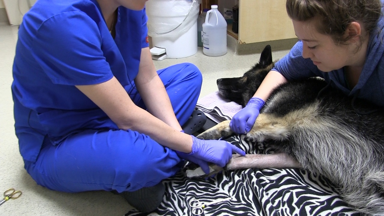
Veterinary suture removal. Suture removal sutures should be removed once there is sufficient healing to prevent the wound reopening. This is usually at 1014 days but healing may take longer in debilitated patients or if there has been patient interference. At 10 to 14 days any external sutures will be removed by your vet and if your dog had absorbable sutures the incision site will be looking less prominent though it can take up to two months for them to be completely absorbed into the body.
Veterinary Dentistry Extraction Introduction The extraction of teeth in the dog and cat require specific skills. In this chapter the basic removal technique for. Absorbable single interrupted sutures spaced no more than 15mm apart.
Advantages Prevents contamination of the site by food and other debris. Suture Removal Scissors are widely used for suture removal. These scissors feature a small hook on one tip that slides under the sutures to slightly lift the suture before cutting and removal.
The hook also holds the suture thus avoid any type of slippage prior to cutting. There are many types of Veterinary Suture Removal Scissors such as Littauer Stitch Scissors Northbent. The suture will be removed in four 12 weeks after the operation making it an ideal choice in some cases where sight or a feeling of pain may prevent your patient from applying pressure on their incision.
Suture the flap to cover the extraction site. The flap may need to be lengthened further by incising the periosteum or undermining the connective tissue. Place sutures 2-3mm apart taking tissue bites of 2-3 mm.
There should be no tension on the flap and the suture line should ideally be placed over bone rather than over the alveolus. November 15 2014 Published. December 12 2014 Abstract The range of available sutures has increased over the last 3 decades.
Wound management is an integral part of daily veterinary practice. All wounds should be considered individually with regards to their most appropriate closure method this is most commonly via suturing. Nurses are able to perform suturing under Schedule 3 of the Veterinary Surgeons Act 1966 when supervised by a veterinary surgeon.
To remove intermittent sutures hold scissors in dominant hand and forceps in non-dominant hand. This allows for dexterity with suture removal. Hold scissors in dominant hand and forceps in non-dominant hand.
Place a sterile 2 x 2 gauze close to the incision site. Suturing of the incision proceeds with simple interrupted non-absorbable suture material using the law of halves. 4-0 to 6-0 suture is used.
Eyelid laxity may also be present with entropion. This is corrected by performing a wedge resection at the lateral canthus either before or after the entropion surgery. Suture reactions can cause morbidity to the patient prolong healing time and increase the treatment costs.
Nonetheless the prognosis is usually very good. When selecting an appropriate suture material one must pay attention to the characteristics of the suture material the condition of the wound environment in question and its healing rate. GerVetUSA produces and sells a wide range of high-quality Suture scissors for veterinarians across the globe.
Our products are used by doctors and surgeons because of their durability and strength. We manufacture the finest quality of Veterinary Suture Removal Scissors such as Littauer Stitch Scissors Northbent Stitch Scissors Spencer Stitch Scissors Health Suture. It is not necessary to use inverting suture patterns nor to do a two-layer closure.
Sutures should be placed 3 to 5mm from the edge of the tissue and around 3mm apart. Minimise how much you handle the edges of your tissue with forceps Figure 4A and use Debakey forceps rather than rat tooth forceps. The submucosa must be included in the closure.
Nonabsorbable sutures are typically chosen to appose tissue that will heal slowly or for tissue in which the suture can be removed non-invasively after healing. Three characteristics of suture materials are important to consider when choosing the proper suture for repair. 1 Initial tensile strength 2 Knot security 3.
Corporate LocationGohad PurDubai Chowk Sialkot. 900 AM 600 PM. Non-absorbable suture materials are either used in areas that allow easy removal after healing eg.
Skin closure or when long term suture strength is required eg. Specific orthopaedic procedures to appose tissue that is expected to heal very slowly or that is under great tension. Absorbable sutures are usually removed in 10-14 days but this may vary by location and situation.
NATURAL SUTURE MATERIALS Natural sutures are made from animal or plant materials. Their protein composition can elicit the most pronounced tissue reaction inflammation of any suture material. Their useful strength in tissue varies.
Thanks for watching the suture tutorial discussing suture removal. In this quick video I demonstration best practices for removing sutures once a wound has. Suture removal Follow up with your veterinarian if suture removal is necessary.
If your new pet has a sutured incision normally the sutures are due for removal in approximately 10-14 days after surgery. The Quill barbed suture is a proven wound closure device that eliminates the need to tie knots during soft tissue approximation. Quill uses a running stitch to hold wound edges together with the strength of an interrupted suture.
The barbs lock the suture in place maintaining tension the length of the wound. Commonly Used Scissors for Suture Removal. There are many types of scissors that vets use to remove sutures.
All of them serve a unique purpose and are ideal for veterinary surgeries. These types of Stitch Removal Scissors are used to remove sutures without causing any damage to the skin. Demonstration of removing sterile suture materialClinical Skills Lab University of Veterinary Medicine Hannover Germany in collaboration with the Universit.